
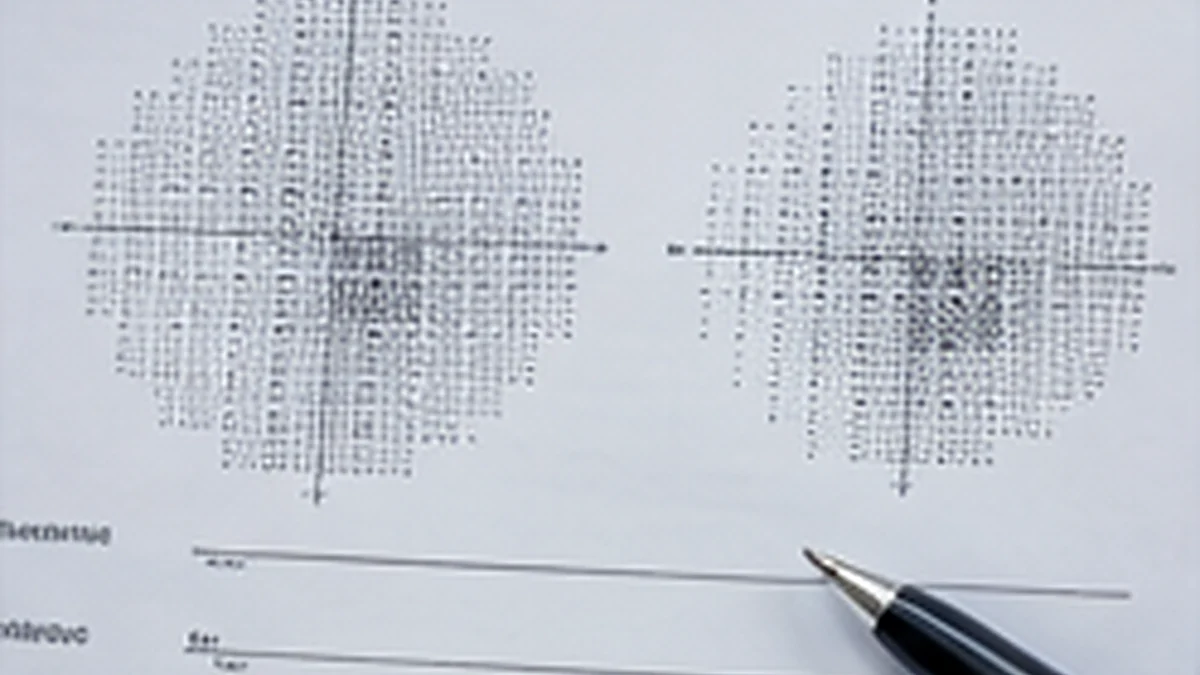
A visual field test printout can look technical, with shaded maps, numbers and reliability measures. These results show how sensitive different parts of the field were during that test. They are not a diagnosis by themselves: an eye care professional interprets the pattern alongside the test quality, symptoms, examination and previous results.
What does a visual field chart show?
Automated perimetry presents lights at different points while you look at a central target. The machine records which targets you noticed and estimates sensitivity across the tested area. The printout may show a visual map and numerical comparisons with expected results for people of a similar age. The exact layout and labels vary between machines and test strategies.
A darker area or a group of missed points can indicate reduced sensitivity, but it does not automatically mean that you have a permanent blind spot or a particular condition. Blinks, loss of fixation, misunderstanding instructions, fatigue, a learning effect, eyelid position, lens correction and other factors can affect a test.
How clinicians check whether a result is reliable
Before interpreting the shape of a defect, the clinician reviews the reliability information for that test. Depending on the equipment, this can include fixation monitoring and responses that suggest a person pressed when no target was shown or missed targets they would usually be expected to see. These measures help identify a test that may need repeating; they do not label a patient as careless or mean that every result is unusable.
Visual fields can vary from one test to another. A repeat may establish a clearer baseline or help determine whether an apparent change is consistent. The College of Optometrists advises that visual field results can be anomalous, and that baseline measurements and ongoing comparisons are useful. Clinicians also compare field findings with other examination results.
Common patterns and what the terms mean
Glaucoma related field changes
Glaucoma can cause characteristic areas of reduced sensitivity, often in the peripheral field. A clinician checks whether the pattern fits with the appearance of the optic nerve and other findings. A visual field abnormality alone does not confirm glaucoma; the result is considered with eye pressure, optic nerve assessment and imaging such as OCT when appropriate.
Hemianopia
Hemianopia means loss of roughly half of the visual field. In homonymous hemianopia, the missing area is on the same side of the visual space in both eyes for example, the left side of the field in each eye. This can be associated with a problem along the visual pathways in the brain, including stroke. A new, sudden field loss needs urgent medical assessment.
Bitemporal hemianopia
Bitemporal hemianopia describes loss of the outer, or temporal, part of the visual field in both eyes. A pattern like this can point the specialist towards the area where the optic nerves meet, called the optic chiasm. There are different possible causes, so the chart cannot identify the cause on its own; prompt clinical assessment and any further tests recommended by the specialist are important.
A bitemporal hemianopia visual field pattern is different from homonymous hemianopia, where the same side of the scene is affected in both eyes. The names describe the location and shape of a field defect, not a complete diagnosis.
Why the clinician compares the chart with other findings
The field pattern is considered alongside what the person notices, visual acuity, examination of the optic nerve and retina, eye pressure and imaging when indicated. The clinician may also compare each eye separately and review older charts. NICE guidance for glaucoma diagnosis recommends standard automated perimetry and repeating the assessment when needed to establish severity.
This is why two charts with a similar looking patch may not have the same explanation. The size, location, consistency, reliability and change over time all matter, as does whether the pattern matches other clinical findings.
Visual field results and driving
A visual field defect may be relevant to driving, but a routine field test should not be used by itself to decide whether someone meets licensing standards. If the DVLA requests an assessment, it specifies the required method; GOV.UK states that a binocular Esterman test is required for DVLA field assessment in the usual process, with other methods considered only in particular circumstances. See the current GOV.UK visual disorders and fitness to drive guidance and our page about DVLA eye tests.
When a visual field change needs urgent attention
If part of your vision suddenly disappears, or a new field defect occurs with face or arm weakness, speech difficulty, confusion, severe headache or problems with balance, seek emergency help. NHS guidance says to call 999 for possible stroke symptoms, including sudden vision loss. Do not wait for a routine visual field appointment.
If you have an existing field defect and a new test shows a possible change, contact the clinician responsible for your care and follow their advice. A changed printout should be reviewed in context rather than self diagnosed.
Frequently asked questions about visual field results
Does an abnormal visual field test mean I have glaucoma?
No. An abnormal result can have several explanations and must be interpreted with the rest of the examination. A clinician may repeat the test and compare it with the optic nerve, eye pressure and imaging.
Can a visual field result be wrong?
A result can be affected by fatigue, fixation, blinking, unfamiliarity with the test or other factors. Reliability information helps the clinician decide whether the chart is useful or whether a repeat is needed.
What is the difference between hemianopia and bitemporal hemianopia?
Hemianopia means loss of about half of the field. Homonymous hemianopia affects the same side of the visual space in both eyes. Bitemporal hemianopia affects the outer side of the field in each eye.
Can I interpret my own visual field printout?
You can read the labels, but the chart should be explained by the clinician who knows why the test was requested and can compare it with your examination and earlier results. Avoid diagnosing yourself from a screenshot.
Discuss visual field results in West London
Specialeyes Eyecare can discuss eye health assessments and explain when visual field findings need further clinical review or referral. We are at 128 Uxbridge Road, Shepherds Bush, London W12 8AA. Call 020 8743 7552 or see our eye health checks in West London.
Clinical review
Clinically reviewed by: Hayder Ali, Qualified Optometrist
Last clinically reviewed:
Further clinical reading
For additional patient information and clinical guidance: