
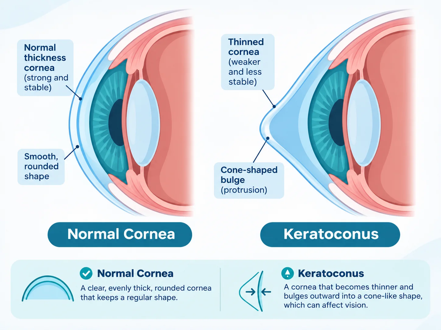
Keratoconus is an eye condition in which the cornea, the clear front surface of the eye, becomes thinner and more irregular in shape. It can develop gradually and may cause short sightedness and irregular astigmatism, leading to blurred or distorted vision. The condition can affect each eye differently, and its course varies from person to person.
What happens to the cornea in keratoconus?
A healthy cornea has a smooth, curved shape that helps focus light. With keratoconus, the cornea gradually thins and steepens, sometimes forming a cone like bulge. This abnormal cornea shape can make it harder for light to focus evenly. People may hear it described as a cone shaped cornea or bulging cornea.
As the shape becomes more irregular, a standard spectacle prescription may become less able to correct all of the distortion. Keratoconus can be mild and stable in some people, while in others the shape changes over time. It does not mean that everyone will need surgery or a corneal transplant.
Common keratoconus symptoms
Keratoconus may cause gradually changing vision. Common signs and symptoms can include:
- Blurred or distorted vision, even with glasses.
- Increasing or irregular astigmatism and changes in short sightedness.
- Frequent changes to a glasses prescription.
- Glare, halos or sensitivity to bright lights.
- Ghost images or difficulty getting a clear, comfortable focus.
These symptoms can have other causes too, so they cannot confirm keratoconus on their own. A change in prescription, especially when one eye changes more than the other, is a reason to arrange an eye examination.
What causes keratoconus?
The exact cause is not fully understood. Keratoconus may be associated with family history and allergic conditions such as eczema, asthma or hay fever. Frequent or vigorous eye rubbing is also linked with keratoconus and may contribute to progression. If your eyes are itchy, seek advice about managing the irritation and try not to rub them.
Keratoconus is often first noticed in the teenage years or early adulthood, but it can be diagnosed at other ages. It may affect both eyes, though usually not to the same degree. No single factor explains every case.
How is keratoconus diagnosed?
An assessment can include a discussion of your symptoms and history, a refraction (spectacle test), examination of the front of the eye with a slit lamp and corneal mapping. Corneal topography or tomography measures the shape of the cornea; some scans also assess its thickness. Comparing measurements over time helps clinicians determine whether the condition is stable or progressing.
At our West London optician, we take time when refracting people with keratoconus. Our team has extensive experience refining spectacle prescriptions for this condition and working to obtain the clearest, most accurate refraction results possible. We can diagnose keratoconus and, where specialist assessment, treatment or ongoing monitoring is needed, refer patients into hospital or private care.
For more detail about the tests involved, see our page about eye tests in Shepherds Bush. Moorfields Eye Hospital explains how refraction, slit lamp examination and corneal mapping may be used in its keratoconus diagnosis and treatment guidance.
Glasses and contact lenses for keratoconus
In earlier or milder keratoconus, glasses or soft contact lenses may provide useful vision. As corneal irregularity increases, glasses may not correct every distortion. A specialist contact lens practitioner may discuss options such as rigid gas permeable (RGP) lenses, hybrid lenses or larger scleral lenses. The most suitable lens depends on the eye, comfort, vision and the person’s daily needs.
Glasses and contact lenses can help correct vision while they are being worn, but they do not stop keratoconus from progressing. If you need glasses, careful refraction is important because the prescription can be complex and may change as the cornea changes. You can also read about our prescription glasses and lenses.
Monitoring and when to seek urgent advice
Once keratoconus is diagnosed, clinicians may recommend repeat eye tests and corneal scans to watch for change. The timing depends on the person’s age, scan results, symptoms and clinical advice. If progression is found, an ophthalmologist can discuss whether treatment such as corneal cross linking is appropriate.
A sudden painful drop in vision can sometimes occur when fluid enters a very thin cornea, a complication known as acute corneal hydrops. Seek urgent eye care advice if your sight suddenly worsens or you develop significant pain. Do not wait for a routine glasses appointment. Moorfields also describes hydrops in its keratoconus patient information.
Frequently asked questions about keratoconus
Is keratoconus an eye disorder?
Yes. Keratoconus is a condition affecting the cornea, the clear front surface of the eye. It changes the cornea’s shape and can cause irregular astigmatism and blurred or distorted vision.
Does everyone with keratoconus need a corneal transplant?
No. Many people manage their vision with glasses or contact lenses, and some may be offered corneal cross linking if the condition is progressing. A corneal transplant is generally considered only in more advanced cases when other options are not providing enough vision or the cornea is significantly scarred.
Can keratoconus be diagnosed during an eye test?
An optometrist can identify and diagnose keratoconus. Assessment may include refraction, examination of the cornea and corneal mapping. Further hospital or private specialist assessment may be arranged where needed.
Eye examinations and keratoconus care in West London
Specialeyes Eyecare is an independent optician at 128 Uxbridge Road, Shepherds Bush, London W12 8AA. We can assess keratoconus, take care with refraction for glasses and refer patients for further treatment or monitoring through hospital and private care pathways.
Call 020 8743 7552 or book an eye test online. For general information about eye examinations and scans, visit our eye test page.
Clinical review
Clinically reviewed by: Hayder Ali, Qualified Optometrist
Last clinically reviewed:
Further clinical reading
For additional patient information and clinical guidance: